Home » Without Label » Diagram Rib Cage With Organs / Thoracic Cage Hd Stock Images Shutterstock - The sternum, commonly known as the breastbone, is a long, narrow flat bone that serves as the keystone of the rib cage and stabilizes the thoracic skeleton.
Diagram Rib Cage With Organs / Thoracic Cage Hd Stock Images Shutterstock - The sternum, commonly known as the breastbone, is a long, narrow flat bone that serves as the keystone of the rib cage and stabilizes the thoracic skeleton.
Diagram Rib Cage With Organs / Thoracic Cage Hd Stock Images Shutterstock - The sternum, commonly known as the breastbone, is a long, narrow flat bone that serves as the keystone of the rib cage and stabilizes the thoracic skeleton.. Anatomy of the elbow 12 photos of the anatomy of the elbow anatomy and biomechanics of the elbow pdf, anatomy elbow fossa, anatomy of elbow dislocation, anatomy of the elbow bones, anatomy of the elbow muscles, human anatomy, anatomy and biomechanics of the elbow pdf, anatomy elbow fossa, anatomy of elbow dislocation. The human rib cage is made up of 12 pairs of ribs, some of which attach to a bony process in the front of the chest called the sternum. The top edge of the manubrium has a depression called the suprasternal or jugular notch. 05.11.2019 · rib cage diagram with organs, find out more about rib cage diagram with organs. An injury to the left rib cage can give rise to severe pain in under the ribs, particularly when the affected person is breathing deeply.
The primary responsibilities of the ribcage involve protecting the thoracic visceral organs, enclosing the thoracic visceral organs, and is included. In this image, you will find thoracic vertebrum, costochondral joint, costal cartilage, costal margin, costal arch, thoracic vertebrum, xiphoid process, xiphisternal joint, body, manubrial sternal joint, manubrium, the sternal notch in it. It includes \(25\) bones, i.e., \(1\) sternum and \(24\) ribs. Anatomy of rib cage muscles : There are seven upper ribs, known as true ribs, which attach to the sternum (breastbone.
How To Draw Rib Cage Youtube from i.ytimg.com The following diagrams are the rib cage diagrams. It is also called rib cage. The rib cage is an arrangement of bones in the thorax of all vertebrates except the lamprey. The lungs are responsible for processing oxygen through the body, while the spleen filters the blood and protects against some bacteria. Two of the most notable organs behind the left side of the rib cage are the left lung and the spleen. It is around 12 to 15 cm long and 4 cm wide, and sits across the lumbar spine. Related posts of rib cage diagram with organs anatomy of human stomach. The human rib cage is made up of 12 paired rib bones;
The human rib cage is made up of 12 paired rib bones;
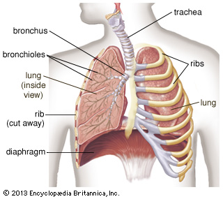
There are 24 ribs in the human body, divided into two sets of 12 curved, flat bones. The ribs partially enclose and protect the chest cavity, where many vital organs (including the heart and the lungs) are located. This organ that is guarded by the rib cage plays a crucial role in aiding digestion. Abdominal cavity chart 14 photos of the abdominal cavity chart abdominal cavity cancer, abdominal cavity contains, abdominal cavity diagram picture, abdominal cavity pain, abdominal cavity quadrants, abdominal cavity regions, air in abdominal cavity, fluid buildup in abdominal cavity, stomach, abdominal cavity cancer. Anatomy of rib cage muscles : It is also called rib cage. The lungs are responsible for processing oxygen through the body, while the spleen filters the blood and protects against some bacteria. Rib cage human anatomy organs. There are 12 pairs of ribs and they attach to the spine. The rib cage is the arrangement of ribs attached to the vertebral column and sternum in the thorax of most vertebrates, that encloses and protects the heart and lungs. There are seven upper ribs, known as true ribs, which attach to the sternum (breastbone. Anatomy of the rib cage diagram. An injury to the left rib cage can give rise to severe pain in under the ribs, particularly when the affected person is breathing deeply.
The sternum, commonly known as the breastbone, is a long, narrow flat bone that serves as the keystone of the rib cage and stabilizes the thoracic skeleton. The rib cage is the arrangement of ribs attached to the vertebral column and sternum in the thorax of most vertebrates, that encloses and protects the heart and lungs. Posted on december 22, 2018december 22, 2018. It encloses and protects the heart and lungs. Rib cage human anatomy organs.
What Is The Function Of The Ribs In The Respiratory System Socratic from useruploads.socratic.org Ribs are mainly of three types: The rib cage is collectively made up of long, curved individual. Diagram of ribs and organs pic of organ in rib cage ribs and internal organ diagram. The human rib cage is made up of 12 paired rib bones; The head of the pancreas resides within the curve of the duodenum, the first section of the small intestine. The area just under the bottom part of the rib cage is the abdomen and not the chest cavity as is commonly thought. They also have a role in ventilation; The rib cage protects vital internal organs.
Rib cage diagram with organs.
There are seven upper ribs, known as true ribs, which attach to the sternum (breastbone. The rib cage is one of the strongest structures in the human body, designed to protect two of the most important organ systems: Ribs are mainly of three types: Rib cage diagram with organs. The last diagram shows how the ribs are connected to the vertebral column or spine. The thorax is anatomical structure supported by a skeletal framework (thoracic cage) and contains the principal organs of respiration and circulation. This diagram depicts human body map of organs with parts and labels. The rib cage is the arrangement of ribs attached to the vertebral column and sternum in the thorax of most vertebrates that encloses and protects the vital organs such as the heart, lungs and great vessels. The rib cage is collectively made up of long, curved individual. Along its inferior margin is a subcostal groove. The rib cage labeled diagram. Anatomy of the elbow 12 photos of the anatomy of the elbow anatomy and biomechanics of the elbow pdf, anatomy elbow fossa, anatomy of elbow dislocation, anatomy of the elbow bones, anatomy of the elbow muscles, human anatomy, anatomy and biomechanics of the elbow pdf, anatomy elbow fossa, anatomy of elbow dislocation. These two organs sit on either side of the body just below the rib cage.
They also have a role in ventilation; Anatomy the rib cage is a bony structure found in the chest (thoracic cavity). 3d illustration of human body skeleton system upper limbs anatomy The following diagrams are the rib cage diagrams. These two organs sit on either side of the body just below the rib cage.
Skeletal Series Part 5 The Human Rib Cage Rib Cage Anatomy Anatomy Bones Human Ribs from i.pinimg.com The kidneys rest against the back muscles, which can make it difficult to tell the difference between kidney pain and back. Two of the most notable organs behind the left side of the rib cage are the left lung and the spleen. Along its inferior margin is a subcostal groove. Abdominal cavity chart 14 photos of the abdominal cavity chart abdominal cavity cancer, abdominal cavity contains, abdominal cavity diagram picture, abdominal cavity pain, abdominal cavity quadrants, abdominal cavity regions, air in abdominal cavity, fluid buildup in abdominal cavity, stomach, abdominal cavity cancer. The rib cage is one of the strongest structures in the human body, designed to protect two of the most important organ systems: It encloses and protects the heart and lungs. Anatomy of the rib cage diagram. There are seven upper ribs, known as true ribs, which attach to the sternum (breastbone.
05.11.2019 · rib cage diagram with organs, find out more about rib cage diagram with organs.
Both the liver and the stomach are located in the lower chest region under the thoracic diaphragm a sheet of muscle at the bottom of the rib cage that separates the chest cavity from the abdominal cavity. 05.11.2019 · rib cage diagram with organs, find out more about rib cage diagram with organs. Rib cage, in vertebrate anatomy, basketlike skeletal structure that forms the chest, or thorax, and is made up of the ribs and their corresponding attachments to the sternum (breastbone) and the vertebral column.the rib cage surrounds the lungs and the heart, serving as an important means of bony protection for these vital organs.in total, the rib cage consists of the 12 thoracic vertebrae and. These two organs sit on either side of the body just below the rib cage. The human rib cage is made up of 12 paired rib bones; How to draw human spine and rib cage diagram. This diagram depicts human body map of organs with parts and labels. It also protects several vital organs of the chest, such as the heart, aorta, vena cava, and. They also have a role in ventilation; Rib cage diagram with organs. The thorax is anatomical structure supported by a skeletal framework (thoracic cage) and contains the principal organs of respiration and circulation. Related posts of rib cage diagram with organs abdominal cavity chart. Anatomy of the elbow 12 photos of the anatomy of the elbow anatomy and biomechanics of the elbow pdf, anatomy elbow fossa, anatomy of elbow dislocation, anatomy of the elbow bones, anatomy of the elbow muscles, human anatomy, anatomy and biomechanics of the elbow pdf, anatomy elbow fossa, anatomy of elbow dislocation.